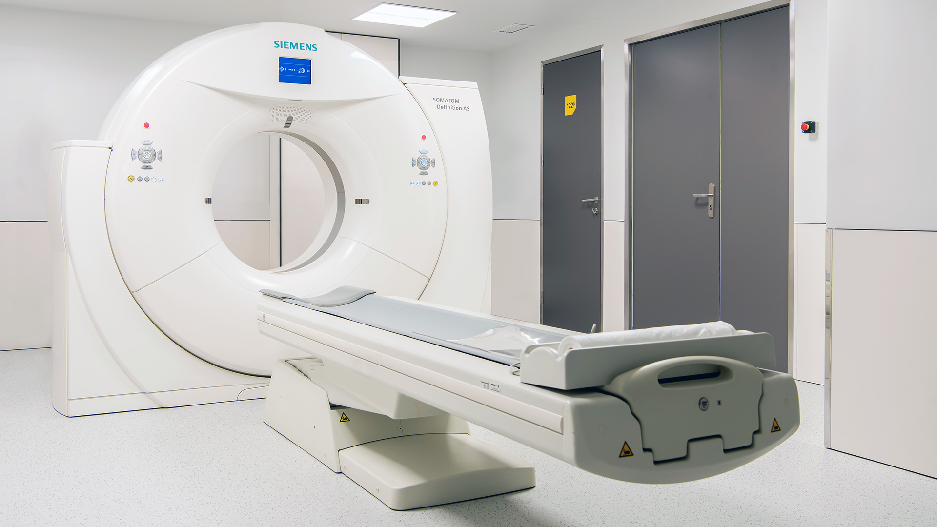
What exactly is Computed Tomography (CT)?

Computed Tomography (CT), also known as a CAT scan, is one of the most effective and widely used diagnostic exams currently available to diagnose and monitor a broad range of diseases. Find out what it is. 

Related Tags

  • CT
  • Imaging
Computed Tomography imaging exam

A CAT scan is a painless and non-invasive exam that enables various structures of the human body to be analysed, by producing axial images that can be reconstructed from multiple angles, providing different perspectives and therefore a more accurate assessment. This is one of the diagnostic exams that currently allows for a more comprehensive, rapid and detailed view of multiple organs. Find out how it is performed and when it is recommended.


What is Computed Tomography?

A CAT scan is an imaging exam that uses X-rays to visualise and assess organs in a specific area of the body. Images are obtained from various angles and in different planes, creating multiple, extremely fine (less than 1 mm) cross-sectional “slices” that are then processed by computer, reconstructing images that enable different structures of the body, such as bones, parenchymal organs and blood vessels, to be viewed and analysed far more effectively than with conventional X-rays.


Therefore, computed tomography provides much more detailed images than conventional X-rays, whose resolution in limited because the different structures of a certain region are superimposed on a single image.


In many cases, CT provides enough information to inform diagnosis and clinical decisions, by allowing disorders to be characterised, narrowing the range of possible diagnoses and providing more accurate indications of any other exams that may be required. In certain cases, a CAT scan can even be sufficient to obtain a final diagnosis, making it possible to avoid other more invasive diagnostic methods.


How is Computed Tomography performed?

CAT scans usually involve the following steps:

1.    Patients should wear a hospital gown and remove all metallic objects. 
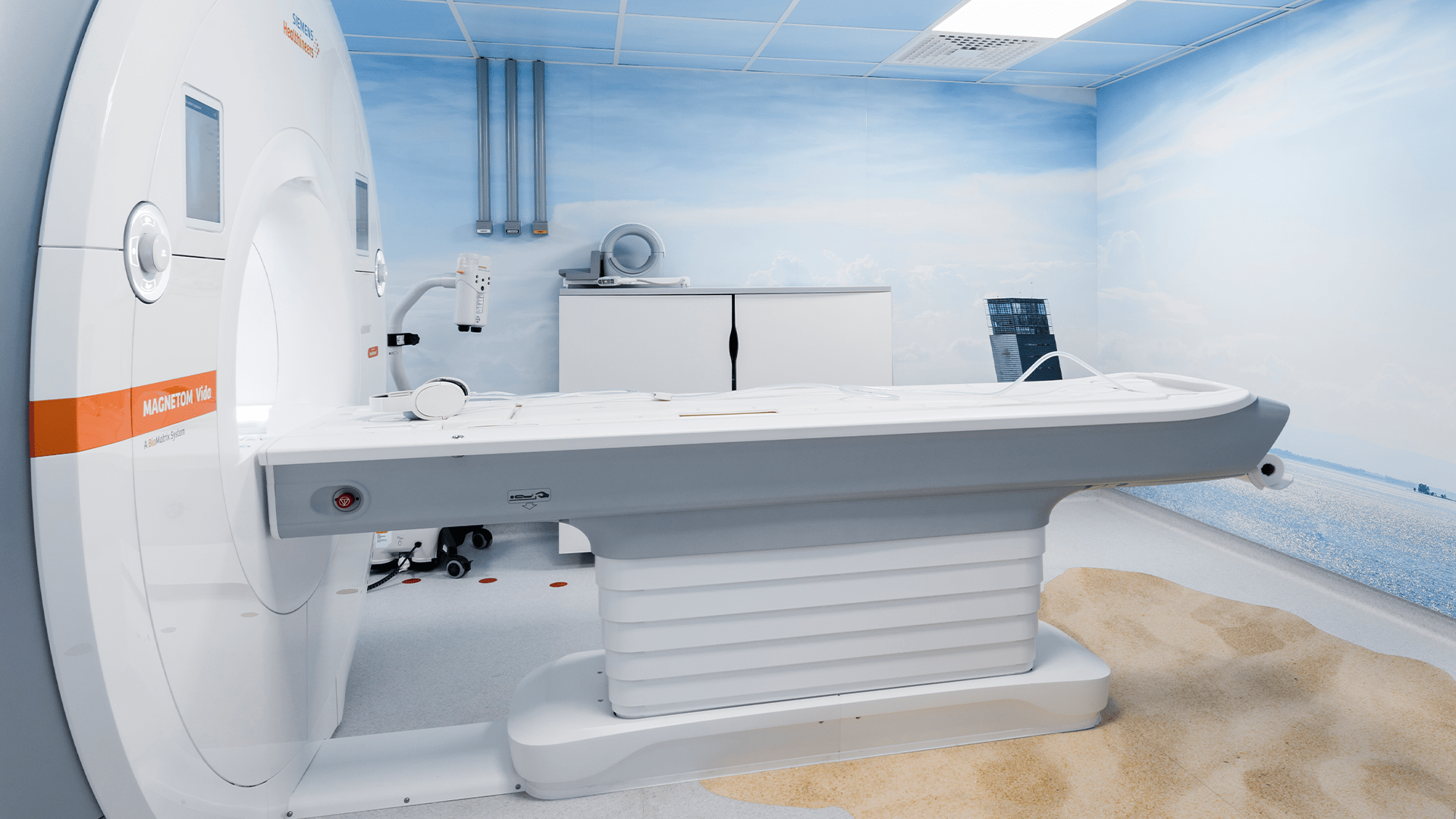
2.   Next, they lie on a table that slides through a donut-shaped scanner.
3.   The scanner rotates around the body as it emits X-rays, obtaining detailed images of the area being studied. To guarantee image quality, it is crucial for the patient to remain still.
4.   The technical team monitors the patient continuously, staying in constant contact and providing instructions whenever necessary. This exam is quick, usually lasting around 10 minutes (duration can vary depending on the study).
5.   The CAT scan can be performed with or without contrast material (usually iodine-based), which is administered orally and/or intravenously. This dye provides a more detailed view of the organs and better characterisation of lesions.
6.   After the exam, the images are analysed by a physician, who produces a report providing an interpretation of the results.

While they are usually safe, contrast media can cause side effects, which in most cases are mild and self-limiting. In certain situations, preventive medication may be required. The use of contrast material should always be assessed by the physician, as it may not be recommended for patients with severe renal insufficiency, pregnant women, or people with a history of moderate to severe allergic reactions.

Which parts of the body can a CT scan be performed on?

CT can be used to assess any part of the body. We describe below the most common types.

  • Cranial Computed Tomography

  • Spinal Computed Tomography

  • Shoulder or Knee Computed Tomography

  • Chest Computed Tomography

  • Abdominal and Pelvic Computed Tomography

  • Cardiac Computed Tomography

What are the limitations and risks of CT?

Although this exam offers high precision and image quality, in many cases CT is not necessarily the first choice, as it uses ionising radiation, thus exposing the body to more radiation than conventional X-rays and, naturally, than other techniques that do not employ ionising radiation, such as ultrasound and magnetic resonance imaging (MRI).

The dose of radiation received by the patient depends on the type of exam and various other factors, although, as a rule, it represents roughly the natural radiation received by any person over a period of several months or years.

Therefore, CT is not recommended for women who are or suspect they may be pregnant, and its use in children should be minimised, as these patients require extra care. Whenever used, if the administration of intravenous contrast is necessary, this carries additional risks, namely of an adverse reaction to the contrast; thus, the risks and benefits of CT should always be weighed for each clinical situation.


What precautions should be taken before undergoing CT? 


The necessary preparation depends on the part of the body to be examined. In most cases, no special preparation is required, but for others certain prior measures are recommended.

For example, in most cranial, spinal, chest and musculoskeletal CT scans, no specific preparation is needed. However, due to the frequent need to administer intravenous contrast, in most CT scans of the neck, heart, abdominal and pelvic regions, the patient should not eat any food in the 4 hours prior to the exam. Furthermore, when the exam is performed on the abdomen or pelvis, it may be necessary to drink a special substance (oral contrast), around 30 minutes before the exam, for a better analysis of the intestine, which may require arriving at the clinic earlier.

Irrespective of these recommendations, you should always take your regular medication with a small amount of water.

As the guidelines for undergoing CT can vary depending on the purpose of the exam, it’s important to strictly follow all instructions provided by the technical and medical team, as some specific precautions may be required at the time. However, it is always necessary to remove all metal objects, such as earrings, watches or bracelets, as these interfere with the quality of the images and consequently affect the result of the exam.


Computed Tomography at Joaquim Chaves Saúde

The Joaquim Chaves Saúde medical clinics feature state-of-the-art CT equipment with 128-slice CT scanning, to provide high quality images that help physicians make a quick and accurate diagnosis, which is essential to determine the best clinical approach for each case. At Joaquim Chaves Saúde you can count on a dedicated imaging team, assisted by the latest technology. 

Clinical Team

We have a team of doctors and health professionals, specialists in various areas, available to give you the support you need.

Luís Curvo Semedo
Medic
Luís Curvo Semedo
Speciality/Service
Radiology
Key areas of expertise
Abdominal and gastrointestinal radiology, Oncological radiology, Urgent/emergency radiology
Healthcare Units
Clínica de Miraflores
Denil Tribovane
Medic
Denil Tribovane
Speciality/Service
Neuroradiology
Key areas of expertise
Neuroradiology, Advanced neuroimaging techniques
Healthcare Units
Clínica de Miraflores, Clínica de Sintra, Clínica de Cascais, Clínica Cirúrgica de Carcavelos
Mário Tavares
Medic
Mário Tavares
Speciality/Service
Radiology
Key areas of expertise
Interventional radiology, Head and neck radiology
Healthcare Units
Clínica de Miraflores

Imaging

Get to know this area of health and find the right care for you.

Book Appointment and Exam

Book an appointment or an exam to receive the best medical care.
Share