Ultrasound is one of the most commonly used tests to evaluate the human body and aid in diagnosis. Discover what it is and when it should be done.
Ultrasound is often the first diagnostic test performed due to its simplicity, safety, and painlessness. It allows for the evaluation of internal structures in virtually any part of the human body. Learn about the types of ultrasound, how it is performed, and the cases in which it is indicated.
What Is Ultrasound and What Is Its Purpose?
Ultrasound is a safe and non-invasive test that generates images of any organ in the body, except for bony structures and organs containing air. It allows visualization of organs, blood vessels, muscles, tendons, and joints. By emitting ultrasound waves and using computer algorithms, the internal structures are reconstructed into an image on a screen, enabling quick and effective visualization.
This test has the great advantage of not using X-rays or magnetic fields. There is no radiation involved, making it suitable for children, pregnant women, and the elderly. Ultrasound assists doctors in diagnosing various diseases and evaluating a patient's response to a specific therapy.
Types of Ultrasounds
There are several types of ultrasounds, depending on the areas of the body being analyzed.
Abdominal Ultrasound
Abdominal ultrasound evaluates the organs located in the abdominal area, including the liver, gallbladder and bile ducts, pancreas, spleen, and major blood vessels. This examination is used to diagnose gallstones and certain liver diseases. Although it has limitations in studying the intestine due to the presence of air, it is used to diagnose acute appendicitis in conjunction with other tests.
Thyroid Ultrasound
Thyroid ultrasound evaluates the thyroid gland located in the neck. It is part of the endocrine system and is essential for hormone production. Therefore, this examination is often requested when thyroid hormone levels are abnormal. It is also used to detect thyroid nodules.
Thoracic Ultrasound
Thoracic ultrasound is less commonly used in this region due to the presence of bony structures that limit visualization. However, due to its quick application, it can detect pleural effusion (fluid in the lungs). It can also be a primary complementary method for diagnosing pneumonia in children.
Renal Ultrasound
Renal ultrasound visualizes the kidneys and ureters, identifying renal tumors and the consequences of diabetes and high blood pressure on the kidneys. It is the first-line examination for diagnosing kidney stones and their complications. It is useful in evaluating renal insufficiency, renal cysts, and kidney infections (pyelonephritis).
Breast Ultrasound
Breast ultrasound is often used as a complementary examination to mammography and aims to detect possible abnormalities in the breast. It allows for the evaluation of the mammary gland without the use of radiation, unlike mammography. It is requested for breast cancer screening in conjunction with mammography and to confirm signs suggestive of malignancy through ultrasound-guided biopsy. It can also be performed individually when a lump is palpated or in the presence of other suspicious symptoms.
Pelvic Ultrasound
Pelvic ultrasound evaluates the pelvic organs in both women and men. It is used to visualize the uterus, fallopian tubes, and ovaries in women, as well as to analyze the prostate in men. In both genders, it enables the assessment of bladder changes, detecting tumors or bladder stones.
Bladder Ultrasound
In this type of ultrasound, the aim is to specifically examine the bladder, making it an important diagnostic tool for benign or malignant tumors, urinary calculi (stones in the urinary system), bladder diverticula, and other bladder pathologies.
Prostate Ultrasound
Prostate ultrasound allows for the evaluation of prostate dimensions, volume, structure, and contours. It is used for diagnosing prostate nodules, confirming the presence of tumors, and detecting calcifications or dilations of the seminal vesicles. It can be performed via suprapubic (through the abdomen) or transrectal approach, with the choice of the most appropriate examination determined by the physician.
Gynecological Ultrasound
Gynecological ultrasound is used to evaluate the uterus, ovaries, endometrium, or bladder in women. It is the preferred examination for diagnosing and monitoring pregnancy. It can be performed via suprapubic approach (placing the probe on the abdomen) or transvaginally (using a high-frequency probe inserted into the vaginal canal). These two approaches can be used individually or together to obtain a more precise diagnosis.
Soft Tissue Ultrasound
Soft tissue ultrasound aims to diagnose localized changes in any superficial anatomical area, allowing for the detection of skin nodules, inguinal hernias, and other abdominal wall hernias. It is used to evaluate the integrity of muscles, tendons, and joint structures.
Cardiac Ultrasound
This type of ultrasound is known as an echocardiogram and is performed to evaluate the heart and major blood vessels. It analyzes the structures of the heart, including heart valves, cardiac chambers, and major cardiac vessels. This examination also assesses cardiac function, analyzing how the heart contracts, how the valves function, and how blood flows. It is essential for diagnosing various types of valvular heart diseases, hypertrophic cardiomyopathy, respiratory insufficiency, aneurysm, or aortic dissection.
How Is Ultrasound Performed?
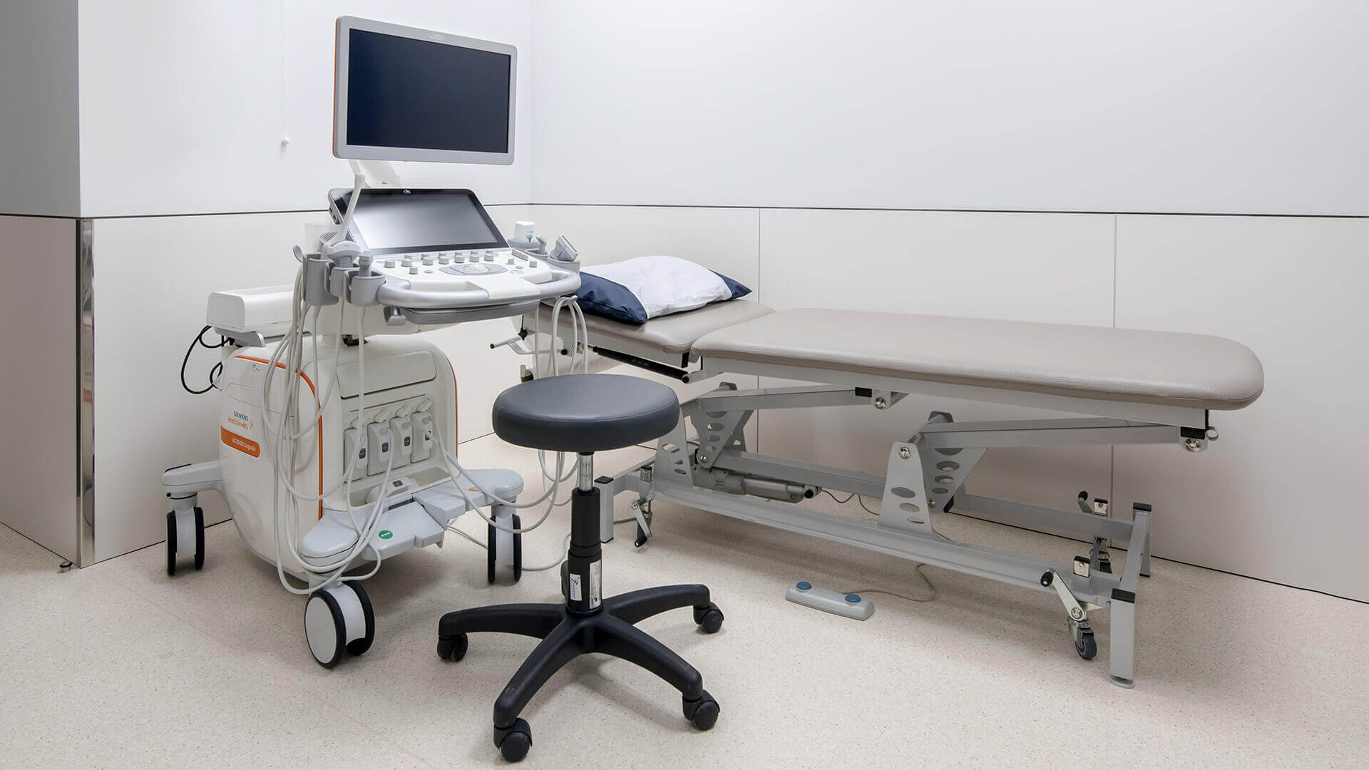
Ultrasound is performed by a specialized doctor who will ask the patient to remove accessories or clothing that interfere with the examination. A conducting gel is applied to the area being analyzed, which allows for the transmission and capture of ultrasound waves. The doctor will then glide a probe over the skin to obtain images. Different types of probes may be used depending on the location of the organ being studied. The images appear immediately on the monitor.
The examination is painless, but the doctor may apply some pressure to gain better visibility of the tissues. During the examination, the images are observed and interpreted, and some of them may be selected for storage and printing. Depending on the area being examined, the ultrasound may take up to 30 minutes, but in most cases, it does not exceed 10 minutes.
Precautions for Ultrasound
Some special precautions are required before undergoing an ultrasound, depending on the area being analyzed. For some examinations, the patient may need to fast, while for others, drinking plenty of water before the ultrasound is necessary. Your doctor will provide all the necessary guidelines specific to the type of ultrasound you will undergo when scheduling the examination.
Limitations and Risks of Ultrasound
Ultrasound carries no risk as it does not involve the use of radiation to obtain images. Therefore, it can be performed on infants, children, and pregnant women without specific protection measures. However, ultrasound cannot evaluate bones or organs containing gas, such as the lungs or intestines. In these cases, clear visualization of the internal structures is not possible, and the healthcare professional may need to resort to other types of examinations such as endoscopy, colonoscopy, computed tomography (CT scan), or magnetic resonance imaging (MRI).
Ultrasound at Joaquim Chaves Saúde
At Joaquim Chaves Saúde medical clinics, we are equipped with the most advanced equipment to provide highly accurate imaging diagnoses. Benefit from the latest technology to undergo your ultrasound in the most comfortable way possible and quickly obtain the most suitable results and treatment for your case. Schedule your ultrasound appointment today.