Obstetric ultrasound is mandatory and essential for monitoring fetal growth. Discover why and when it should be done, as well as how to prepare.
According to the guidelines of the General Directorate of Health, obstetric ultrasound should be performed at least three different times during pregnancy. The objective is to monitor the fetus's development, allowing for early detection of possible gestational complications. Learn what it entails and how to prepare.
What is Obstetric Ultrasound?
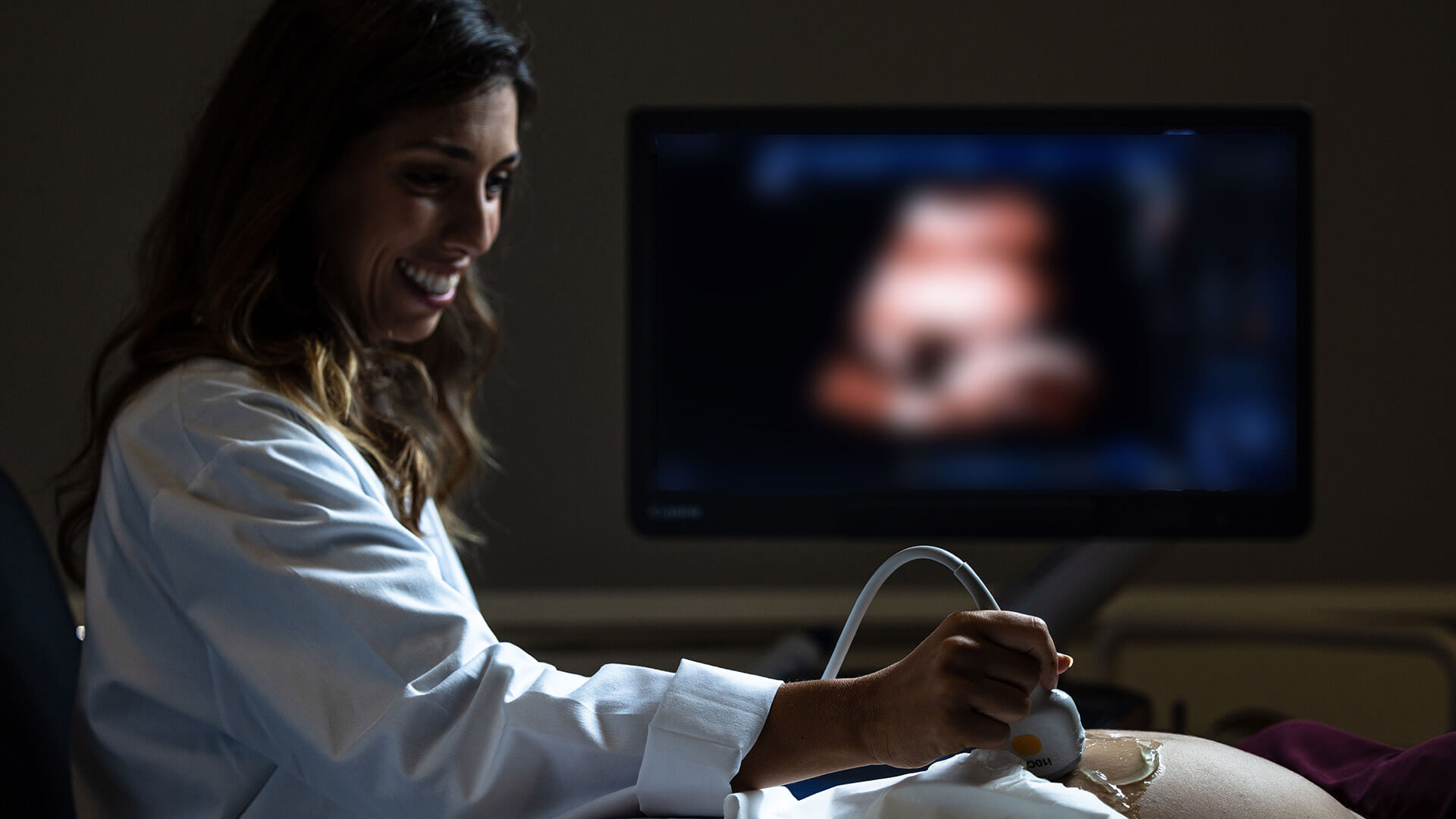
Obstetric ultrasound is an imaging exam that allows visualization of the fetus inside the maternal womb, thereby assessing the baby's health throughout pregnancy. This exam also enables visualization of the placenta, amniotic fluid, umbilical cord, and maternal pelvic structures.
The technique can be performed vaginally (transvaginal ultrasound) or abdominally, depending on the gestational age and accessibility to the fetus. It is a safe and harmless exam for both the baby and the mother since it only uses ultrasound to obtain images.
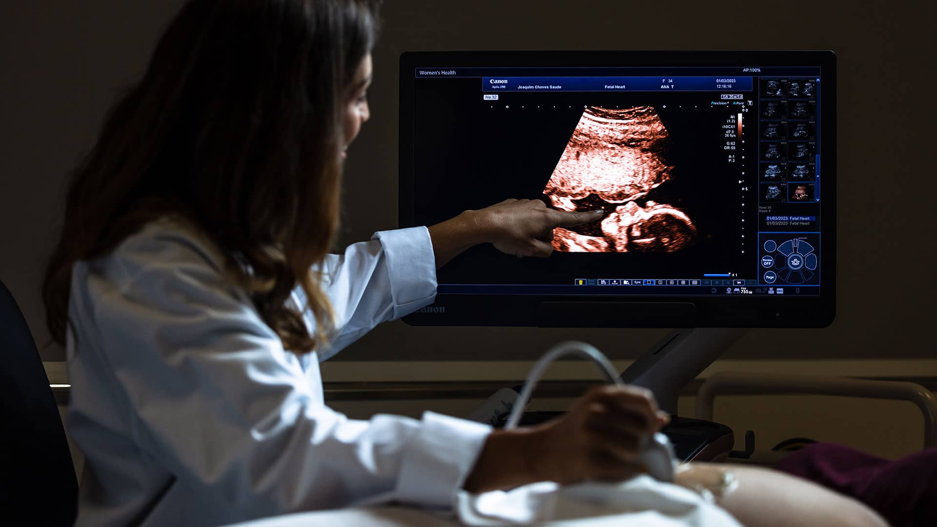
Currently, 3D and 4D obstetric ultrasounds can be performed to visualize the baby in three dimensions and real-time. Although they are not part of routine pregnancy surveillance, they can be important for documenting some morphological changes. However, they are primarily sought after as emotional comfort for parents and should only be performed after excluding anomalies.
Obstetric ultrasound can be performed at any time and repeated as necessary throughout pregnancy, but there are three crucial moments when its execution is crucial and therefore mandatory.
When Should Obstetric Ultrasound Be Performed?
According to the General Directorate of Health and the Ultrasound Technical Committee, there are three mandatory obstetric ultrasounds, which we will now describe.
1. First Trimester Obstetric Ultrasound
The first trimester obstetric ultrasound is performed between 11 and 14 weeks of pregnancy and has the main objectives of determining gestational age (and consequently, the estimated due date) and detecting possible fetal anomalies.
Gestational age is determined with high reliability by evaluating the crown-rump length, biparietal diameter, and head circumference. Fetal viability is assessed based on various factors, including heart rate, external and internal fetal anatomy, chromosomal screening, and evaluation of several ultrasound markers.
This way, it is possible to early screen for chromosomal abnormalities, heart conditions, and some genetic diseases with an accuracy rate of approximately 70%. Along with the mother's age and biochemical screening, obstetric ultrasound can detect Down syndrome (and other chromosomal abnormalities) with a precision level of up to 95%.
2. Morphological Obstetric Ultrasound
The second trimester morphological obstetric ultrasound is performed between 18 and 24 weeks of pregnancy and is done abdominally. At this stage, due to the increased size of the baby, it is possible to observe the fetal anatomy more accurately and detect any physical anomalies.
During this obstetric ultrasound, the Doppler measurement of the uterine arteries is also performed to determine the risk of pre-eclampsia (hypertension associated with protein in the urine). In some cases, it is already possible to determine the baby's gender at this stage.
3. Third Trimester Obstetric Ultrasound
The third trimester obstetric ultrasound is performed between 28 and 32 weeks of pregnancy and is used to detect fetal malformations that only emerge at this stage of gestation (late-onset anomalies). Due to the larger size and better positioning of the fetus, it is possible to evaluate certain previously inaccessible structures.
This obstetric ultrasound assesses the well-being and growth of the baby through biometrics, estimated weight, biophysical profile, and Doppler evaluation of fetal compartments. In situations where there is a risk of fetal growth restriction, this exam can be scheduled for 28 weeks.
In summary, these three obstetric ultrasounds are complementary and of great importance in monitoring the baby. Omitting any of them implies missing the opportunity to detect fetal pathologies at the appropriate time.
When any of the mandatory obstetric ultrasounds raise clinical suspicions, it may be necessary to resort to other complementary diagnostic tests, such as obstetric ultrasound with flowmetry (to evaluate blood flow in the umbilical cord, fetus, and placenta), amniocentesis, chorionic villus sampling, fetal echocardiogram, fetal magnetic resonance imaging, among others that the Obstetrician considers essential.
Additionally, in high-risk pregnancies, it may be necessary to perform obstetric ultrasounds more frequently, with the decision being solely up to the Obstetrician.
What are the limitations of obstetric ultrasound?
Despite scientific innovation and laboratory technological development, obstetric ultrasound still has some limitations. Not all fetal malformations can be detected during pregnancy. The accuracy of this examination depends on several factors, including:
- Gestational age
- Fetus (fetal position, amniotic fluid, etc.)
- Type of anomaly
- Maternal biotype
Therefore, obstetric ultrasound should be performed by a specialist in Gynecology and Obstetrics with recognized expertise in ultrasound. Likewise, parents are always informed that ultrasound does not guarantee 100% detection of malformations.
How to prepare for obstetric ultrasound?
Undergoing an obstetric ultrasound does not require any special preparation or prior care. It is only recommended not to apply moisturizing cream to the abdominal area on the day of the exam and to remove any piercings in that area. The mother should always bring her Pregnancy Health Booklet, previous ultrasounds, and lab tests.
The duration of an obstetric ultrasound varies depending on the type of exam, fetal position, and maternal biology. However, on average, an obstetric ultrasound can take between 30 and 60 minutes. It is a painless and safe exam, without any risk or complications for the baby or the mother.
Joaquim Chaves Saúde, for a safe and peaceful pregnancy
We understand how important it is to fully experience pregnancy. Count on Joaquim Chaves Saúde to feel at home and take care of yourself and your baby's health. Benefit from the most advanced diagnostic technology and an experienced team. Schedule your obstetric ultrasound and experience pregnancy with more peace of mind.