Just by having a 3D ultrasound, you can see the baby. Discover what this examination is, the risks involved, and the best time to undergo it.
A 3D ultrasound allows for a highly realistic visualization of the fetus while still in the womb. However, before undergoing this examination, it's important to know when it should be done and if there are any associated risks to ensure that you're doing everything possible to keep the baby safe. Get all your questions clarified here.
What is a 3D Ultrasound?
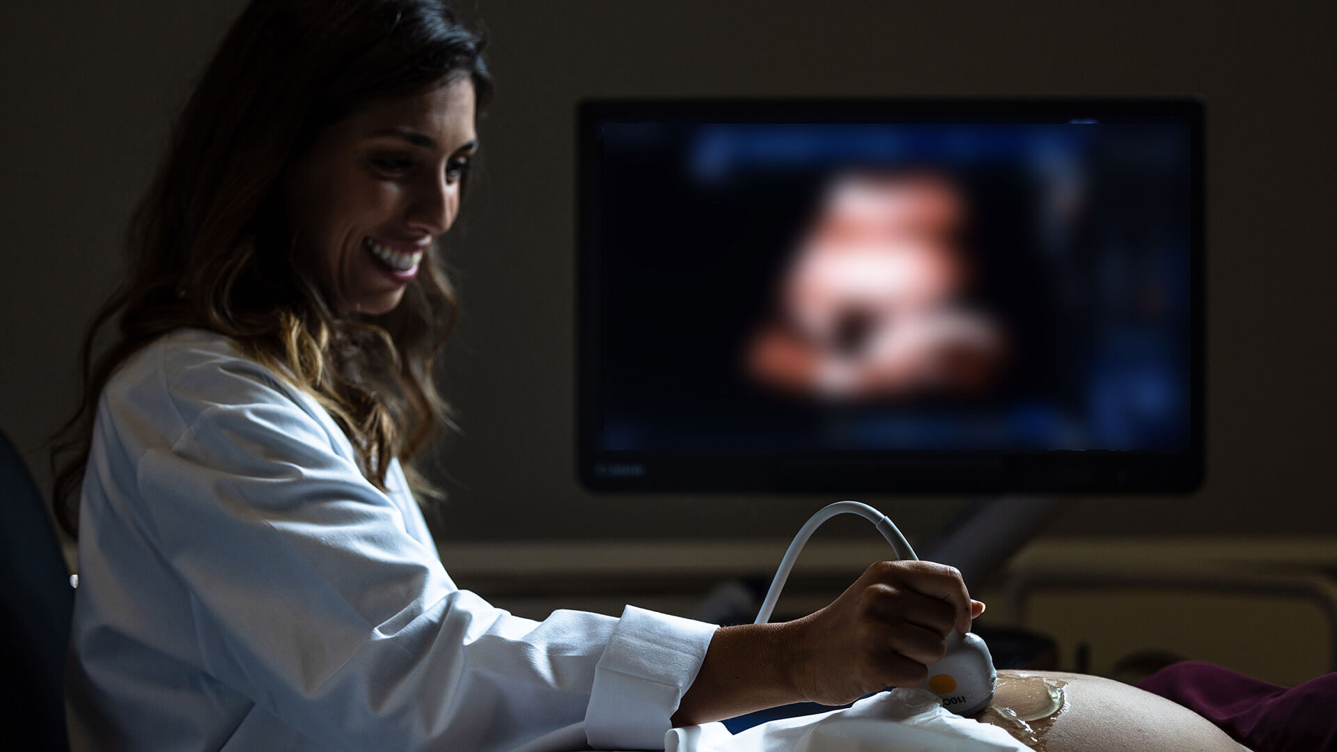
As the name suggests, a 3D ultrasound is a type of ultrasound that enables the observation of the fetus in three dimensions with a high level of realism. For this reason, the 3D ultrasound is often referred to as an emotional ultrasound. It is an innovative procedure that utilizes ultrasound waves without any form of radiation. As a result, it is a completely safe, painless, and comfortable examination. After the procedure, the doctor can record the images in digital format and make them available to the parents. It's important not to confuse this procedure with a 4D ultrasound. While a 3D ultrasound provides static images in three dimensions, a 4D ultrasound generates a real-time video, allowing the observation of the baby's movements, smiles, or yawns.
What is the purpose of a 3D Ultrasound?
A 3D ultrasound is not a diagnostic or obstetric examination, and it does not replace the mandatory 2D conventional ultrasounds used for pregnancy monitoring. It is solely an emotional technique that allows for early contact between parents and their unborn child.
Therefore, 3D ultrasounds are often requested by parents who want to see the baby's face, hands, feet, and real expressions. By "meeting" their child before birth, a 3D ultrasound enhances a pre-existing connection.
However, there are specific cases in which a doctor may recommend a 3D ultrasound. When a more detailed and in-depth evaluation of the fetal anatomy is required, this examination enables the diagnosis of malformations, particularly in external organs.
Additionally, when used in conjunction with a conventional 2D ultrasound, a 3D ultrasound can play an important role in the early diagnosis of cleft lip, cleft palate, spina bifida, and other craniofacial and skeletal malformations. It can also help identify certain chromosomal or genetic diseases due to its ability to visualize an infinite number of planes.
What are the limitations of a 3D Ultrasound?
A 3D ultrasound is a safe and harmless procedure for both the fetus and the mother. Since it only employs ultrasound waves without any radiation or X-rays, it can be performed throughout pregnancy without any risk and can be repeated as desired by the couple.
However, like any examination, a 3D ultrasound should only be conducted by an obstetrician or under their supervision to ensure that the procedure is performed correctly and safely. There are also some factors to consider when undergoing a 3D ultrasound. The success of this examination is not always guaranteed and depends on factors such as the position of the baby, gestational age, amount of amniotic fluid, or the position of the umbilical cord.
When these factors are not optimal, the image may appear distorted, and the visualization may not be optimal. In such cases, it may be necessary to repeat the 3D ultrasound to obtain images of the desired quality.
How is a 3D Ultrasound performed?
A 3D ultrasound is performed in the same way as a conventional 2D ultrasound, with the only difference being the sophistication level of the equipment. The pregnant woman lies down as still as possible, and the doctor applies a conducting gel to the area to be analyzed. Then, a probe is moved over the skin, and the images appear immediately on the monitor.
The average duration of a 3D ultrasound ranges from 30 to 60 minutes, depending on the position of the fetus and the amount of amniotic fluid. The examination does not cause any pain or discomfort for the mother or the baby.
When should you have a 3D Ultrasound?
Although a 3D ultrasound can be performed at any time during pregnancy and repeated as desired, the best time to undergo it is from 25 weeks onward, when the baby's features are well-formed and the face can be visualized. From 32 weeks onward, the obtained images offer an impressive level of realism, almost identical to what the baby will look like at birth. However, obtaining good images can be more challenging due to the limited space the baby has at this stage.
How to prepare for a 3D Ultrasound?
Preparation for a 3D ultrasound does not require any specific measures. However, it is recommended that the pregnant woman avoids consuming coffee, increases water intake, and pays special attention to her diet in the days leading up to the examination. Additionally, the most suitable timing should be considered to meet the parents' expectations.
3D Ultrasound at Joaquim Chaves Saúde
You can undergo your 3D ultrasound at Joaquim Chaves Saúde and benefit from the most advanced technology for that long-awaited first contact with your baby. Schedule your ultrasound and experience your pregnancy with full intensity.