An echocardiogram is a simple, non-invasive exam that is essential to determine if the heart is functioning correctly. Discover how it’s done and what precautions to take.
What is an echocardiogram?
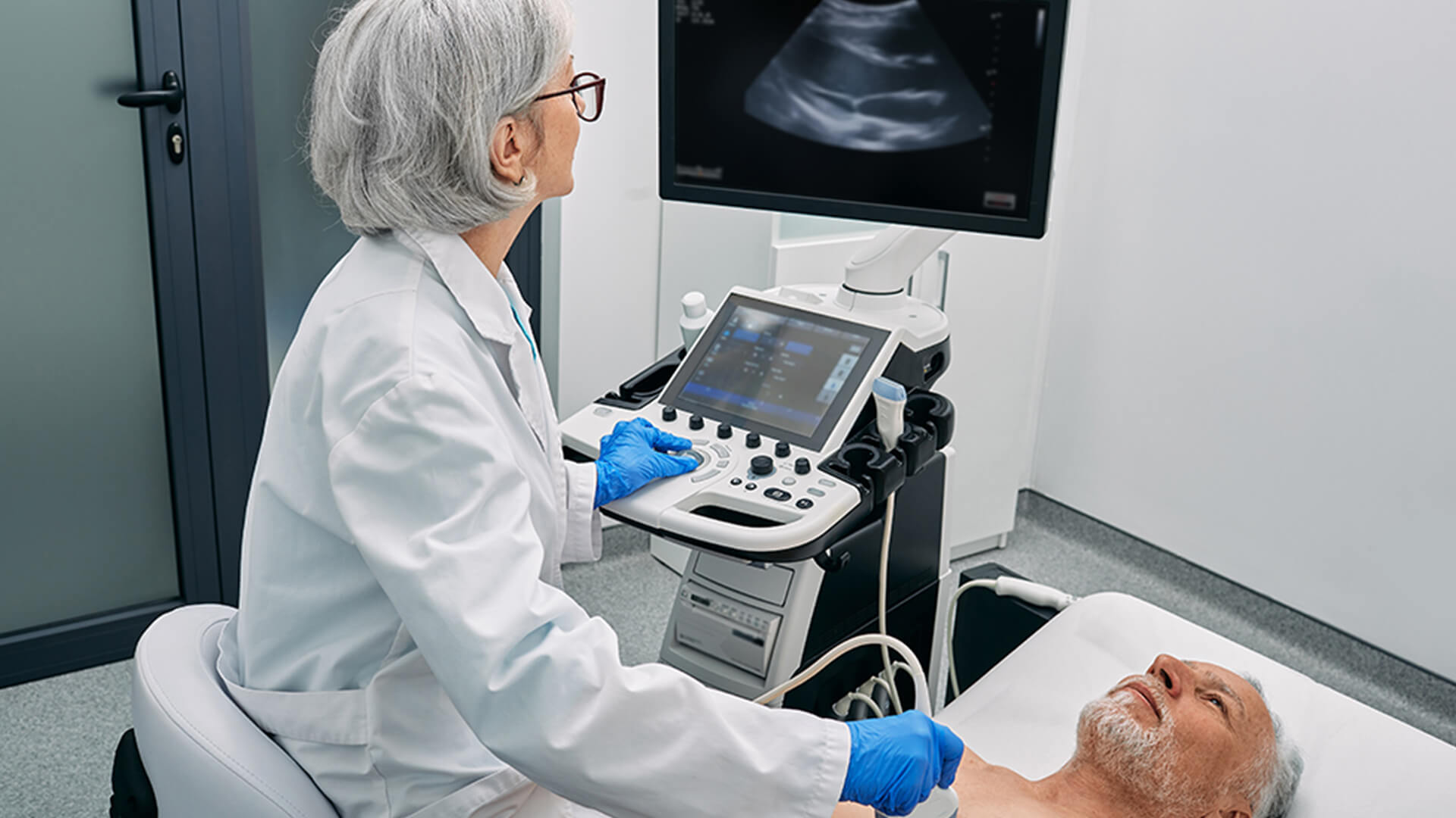
An echocardiogram is an imaging exam that uses ultrasounds to produce real-time images of the heart.
Thanks to this non-invasive technique, physicians can assess the heart’s structures and function – analysing the size of the cavities, pumping function, blood flow and even potential heart valve defects. This exam is fundamental to diagnose and monitor various heart conditions – and it’s quick, painless and radiation-free.
What is an echocardiogram for?
Echocardiograms help detect and monitor various heart conditions.
Diagnose heart disease
This exam is frequently used to identify disorders like heart failure, heart valve disorders, cardiomyopathies and congenital defects.
Assess heart function
Echocardiograms measure the heart’s capacity to pump blood, and help detect signs of weakness in the heart muscle or other disorders that may compromise blood circulation.
Monitor pre-existing conditions
For people with a diagnosed heart disease, this exam is an essential tool to monitor the condition’s progress and adjust treatment when necessary.
Detect clots or swelling
In addition to the heart’s structures, echocardiograms can identify clots, infections and swelling in the heart, factors that can represent serious health risks.
What are the vs?
There are various types of echocardiograms and each one is prescribed by a cardiologist, according to each patient’s needs. Although all types use ultrasound, how they are performed and the information they provide can vary.
Diagnose heart disease
This exam is frequently used to identify disorders like heart failure, heart valve disorders, cardiomyopathies and congenital defects.
There are three modes of transthoracic echocardiograms:
Two-dimensional TTE (M & 2D) - Basic:
The M-Mode provides one-dimensional images to take precise measurements of the heart cavities and wall thickness.
The 2D-Mode creates images of the heart in motion, aiding anatomical and functional assessment.
Doppler TTE - Intermediate:
Measures the speed and direction of blood flow, and is essential to assess heart murmurs, measure pressure gradients and identify valve stenosis (constriction) or failure.
Tissue Doppler TTE - Complete:
Analyses myocardial movement, allowing early detection of cardiac dysfunction, especially myocardial disorders.
Transesophageal echocardiogram (TEE)
In this exam, the probe is inserted into the oesophagus through the mouth /throat, to obtain more detailed images of the heart, particularly the valves. This method is used when the transthoracic exam does not provide enough information. The procedure is performed under mild sedation, takes approximately 20 to 30 minutes, and may require a short recovery period after the exam.
Exercise stress echocardiogram
This type of echocardiogram analyses the heart under exertion, whether through physical exercise or with drugs that simulate exertion. This test is useful to diagnose coronary artery disease and evaluate blood flow.
Firstly, a transthoracic echocardiogram is performed at rest. Next, the patient exercises on a treadmill or bicycle, or medication is administered to simulate exertion. During and after exertion, new images are made of the heart to measure the impact of stress on the heart.
Foetal echocardiogram
This type of echocardiogram assesses the baby’s heart structures and function while still in the womb, to detect congenital defects, arrhythmias and other disorders, before birth. The exam is non-invasive, painless and has no impact on the pregnancy. The results are analysed by a paediatric cardiologist.
The test is usually performed between 18 and 24 weeks of pregnancy, the ideal phase to obtain precise images of the foetus’ heart. The exam is carried out by applying gel and using an ultrasound transducer on the pregnant woman’s abdomen. It takes between 30 and 60 minutes, depending on the baby’s position and complexity of the analysis.
Doppler technology
Doppler technology uses soundwaves to measure the direction and speed of blood flow through the heart and arteries. The flow is shown in colour-coded images that help identify constriction, regurgitation or blood circulation disorders. This procedure can be applied in conjunction with transthoracic, transesophageal, exercise stress or foetal echocardiography.
Limitations of echocardiograms: what you should know
Although these exams are essential to analyse the heart, echocardiograms present some limitations. In some cases, the quality of the images can be affected by the patient’s anatomy, such as people with hyperinflated lungs or obesity, which make it difficult to clearly visualize the heart’s structures. In these cases, it may be necessary to perform a transesophageal echocardiogram to obtain more precise images.
Furthermore, even with Doppler technology, echocardiograms may not provide all the answers on coronary circulation. It is often necessary to complement this test with other exams, such as cardiac magnetic resonance imaging (MRI), computed tomography angiography or myocardial perfusion scintigraphy. Despite these limitations, echocardiography remains one of the safest, quickest and most accessible methods to evaluate the heart and determine the next steps in diagnosis and treatment.