Computed Tomography (CT) or CT scan is one of the most common and effective tests for diagnosing and monitoring various types of diseases. Learn what it is and how it is performed.
In a painless and non-invasive manner, CT allows the investigation of various structures in the human body from multiple angles and in multiple slices. As a result, it is one of the most comprehensive and detailed diagnostic tests available today. Discover how it is performed and in which cases it is recommended.
What is Computed Tomography?
Computed Tomography (CT), also known as CT scan, is an imaging test that uses X-rays to visualize the organ or region of the body being analyzed. The images are obtained from various angles and different planes, in slices smaller than 1 mm, which are later processed by a computer to reconstruct an image that allows for the distinction of bone structures, organs, and blood vessels in a superior way compared to conventional X-rays. Therefore, computed tomography provides a clearer and more detailed image than conventional X-rays, which are limited by the overlapping of internal structures.
This test is non-invasive and does not cause any pain. It is often sufficient to reach a definitive diagnosis, eliminating the need for more aggressive and painful procedures.
What types of Computed Tomography are there?
CT scans can be performed with or without the use of contrast. Contrast agents may be recommended when a sharper visualization of certain structures is required.
However, contrast is contraindicated in patients with severe renal insufficiency, pregnant women, and patients with allergies to contrast agents. The use of contrast may cause various side effects or reactions, which can be properly controlled with prophylactic medication. In these cases, it is crucial to strictly follow all the doctor's recommendations.
The administration of contrast is done in the presence of a qualified nurse and with the support of emergency equipment.
In which body areas can Computed Tomography be applied?
CT scans can be performed in any body area. Let's describe the most common types.
Computed Tomography of the Skull
As the name suggests, this exam is performed on the skull and is indicated to evaluate head injuries, headaches, motor activity changes, sensory alterations, or seizures. It is a diagnostic tool for ischemic or hemorrhagic stroke, hydrocephalus, aneurysm, or infection. It is also very useful for monitoring oncological diseases.
Computed Tomography of the Spine
CT scan of the spine allows for the evaluation of the entire spine, including the cervical, thoracic, lumbar, and sacrococcygeal regions. It can be used when patients present complaints of pain, sensory alterations, or weakness in the arms or legs, with or without previous trauma. It allows for the diagnosis of fractures, congenital vertebral abnormalities, herniated discs, and degenerative diseases such as arthritis or spondylosis. It is also a way to assess the response to treatment of bone metastases or primary tumors in these areas.
Computed Tomography of the Knee
This type of computed tomography is indicated to clarify alterations observed in the physical examination of the knee or in other performed tests (such as X-ray or ultrasound). It can be applied to any structure of the knee, but it is less precise in soft tissues (muscles, tendons, menisci, and cartilage). In pediatric patients, it helps study femoropatellar instability, a condition in which the patella dislocates from its normal position.
Computed Tomography of the Shoulder
CT scan of the shoulder is performed to clarify alterations that are first diagnosed through an X-ray examination, being particularly useful in evaluating the bone structures that make up the shoulder, allowing for the precise diagnosis of fractures and dislocations. It is indicated for trauma or degeneration due to excessive use of this joint. Sports activities and work accidents are often responsible for these injuries. This exam assists in the diagnosis of pathologies involving the muscular and tendinous structures of the shoulder, identified through ultrasound or through a physical examination performed by your doctor.
Thoracic Computed Tomography
Thoracic Computed Tomography (CT), also known as Chest CT, is an examination that thoroughly evaluates the structures of the chest. It is superior to X-rays in many aspects, particularly in the diagnosis of lung diseases, thoracic vascular diseases, and thoracic traumas. It is used to monitor the progression of severe respiratory infections, including Covid-19 infection and Long-Covid syndrome. It can be used in the presence of respiratory difficulty (dyspnea), wheezing, fatigue, or chest pain.
Abdominal and Pelvic Computed Tomography
This examination is indicated for studying the abdominal organs. It allows for the evaluation of the intestines, liver, spleen, kidneys, and abdominal blood vessels, especially when combined with intravenous contrast. It also allows for the evaluation of the uterus, ovaries, uterine tubes, and bladder. When combined with oral contrast, it provides superior imaging of the digestive tract (stomach, small intestine, and colon).
This examination can assist in the diagnosis of acute appendicitis, pancreatitis, Crohn's disease, uterine fibroids, ovarian cysts, kidney stones, and other pathologies. In an oncological context, along with other examinations, it enables the diagnosis and staging of tumors in any abdominal organ.
Cardiac Computed Tomography
This type of CT scan is mainly used to evaluate the coronary arteries and the risk of acute myocardial infarction. When there is a high amount of calcium in these arteries (atherosclerosis), there is reduced blood flow, causing angina in the early stages and myocardial infarction in more advanced stages.
Cardiac computed tomography is also useful for detecting structural abnormalities of the heart, heart valves, pericardium (the membrane that surrounds it), and blood vessels originating from the heart.
What are the limitations and risks of CT?
Although CT is a highly precise and high-quality imaging examination, it is rarely the first option as it exposes the body to more radiation than conventional X-rays. Typically, the radiation dose received during a CT scan is equivalent to three years of exposure to natural background radiation. For this reason, CT is not recommended for pregnant women or women suspected of being pregnant.
Additionally, specific precautions need to be taken for children. If intravenous contrast is used, it may be contraindicated in some diabetic patients, patients with chronic kidney disease, contrast allergies, or asthma. In these cases, the doctor will determine if the examination is viable and, if so, the specific precautions to be taken.
What precautions should be taken during a CT scan?
The precautions vary depending on the area of the body being examined. In most cases, no special preparation is required, but in others, some prior measures may be recommended. For example, for chest, head, and neck CT scans, the patient should not consume any food for 4 hours before the examination, although they can take their regular medication with a small amount of water.
When the examination involves the abdomen and pelvis, it may be necessary to consume a special liquid (oral contrast) 30 minutes before the examination to improve visualization of the intestines.
In this case, the patient will be asked to arrive earlier. It is important to strictly follow all the instructions given by the doctor, as CT scan guidelines vary depending on the purpose of the examination.
However, it is always necessary to remove any metal objects, such as earrings, watches, or bracelets, as they can interfere with the scan results.
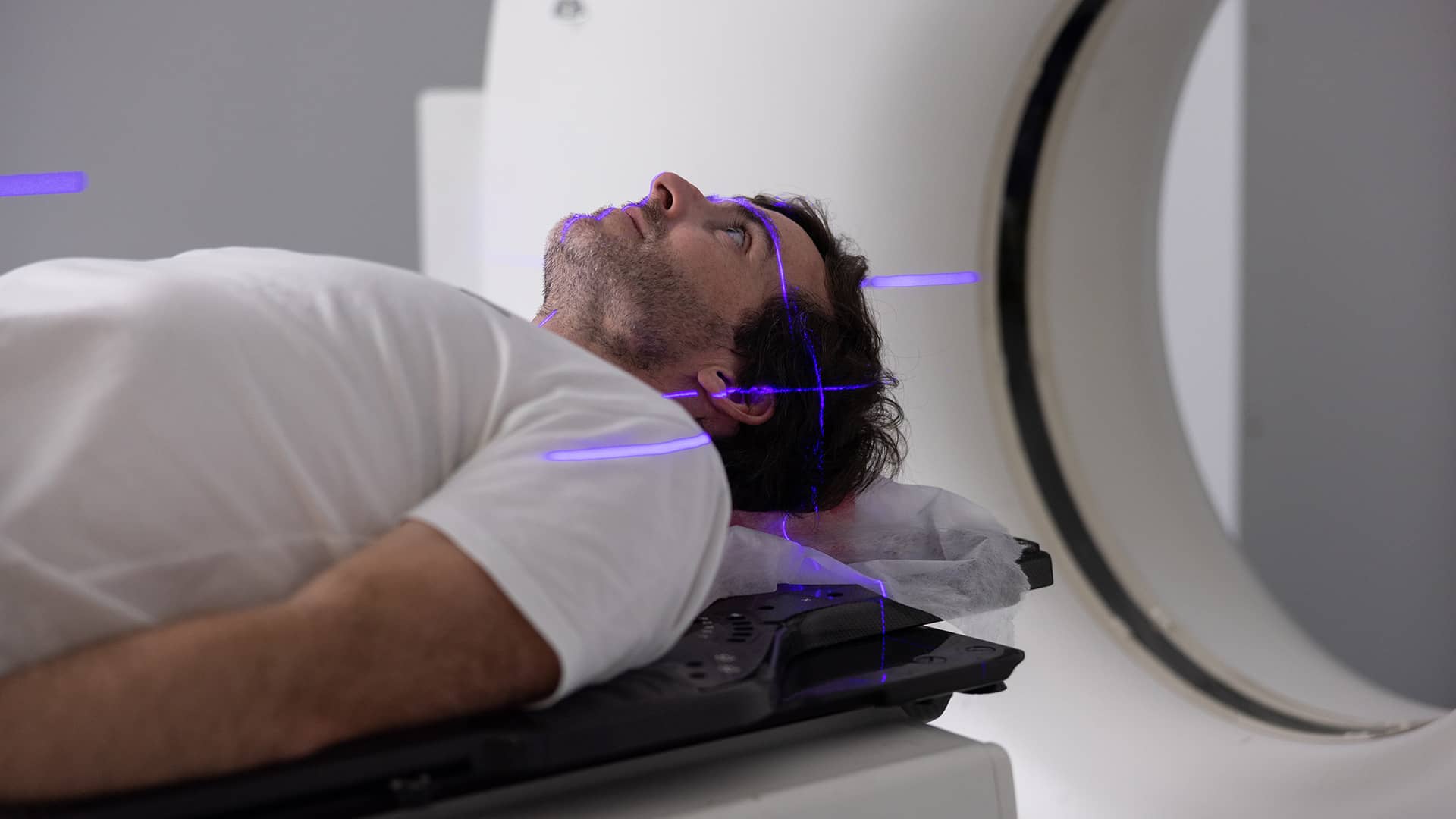
How is CT performed?
Before the examination, the patient is asked to put on a gown and remove all metal objects. Then, they lie on their back on a table and remain as still as possible because any movement can affect the obtained image. The table moves into the CT scanner, which emits X-rays and rotates around the patient to produce the images.
During the process, the healthcare professionals can see the patient through a window and maintain constant communication, providing instructions whenever necessary and interrupting the examination if the patient desires. CT scanning is performed with an open machine, unlike magnetic resonance imaging. On average, the examination lasts about 10 minutes. If the CT scan involves intravenous contrast, the patient may feel warmth in the arm or a metallic taste in the throat.
These are expected reactions, slightly discomforting, and naturally disappear. Subsequently, the physician interprets the obtained images and prepares a report describing any observed abnormalities. The administration of intravenous contrast is done in the presence of a nurse and with appropriate emergency equipment.
Computed Tomography at Joaquim Chaves Saúde
At Joaquim Chaves Saúde medical clinics, we provide state-of-the-art CT equipment, such as 128-slice CT, to achieve a fast and accurate diagnosis, essential for determining the most appropriate therapeutic approach for each case. You can count on a dedicated team supported by the most advanced equipment.