X-ray, also known as conventional radiography, is one of the most commonly used medical examinations. Find out in which situations it should be done and the associated risks.
X-ray was discovered by accident in 1895 in a laboratory in Germany, but it remains to this day the most used and effective examination for quickly identifying changes in the structure of bones and organs. Learn how it works, when to do it, and if there are any associated risks.
What is X-ray?
X-ray is a type of radiation used in various medical examinations to produce images of internal body structures. The goal is to diagnose changes, fractures, various diseases, different types of tumors, or even the presence of foreign bodies.
It is one of the most common and oldest medical imaging techniques. However, its operation is very simple: the emitted radiation is absorbed or reflected by the structures being evaluated. Different tissues absorb different amounts of radiation, resulting in images in various shades of black and white. For example, in these examinations, bones appear white because calcium has a higher capacity to absorb radiation. On the other hand, fatty tissues do not absorb as much, so they appear less white. Lungs, on the other hand, appear black because air practically does not absorb radiation.
In some cases, it may be useful to introduce a contrast agent into the body, intravenously or orally, to allow better visualization of certain structures. Examples include the evaluation of blood vessels and the digestive tract, including the bile ducts. Due to their nature, the obtained images are static. They can be recorded on film, but with technological advancements, this support has fallen into disuse. Currently, they are recorded in digital format.
What is X-ray used for?
X-ray can be used to examine most areas of the body. It is most commonly used to evaluate bones and joints, but its application goes beyond that. X-ray helps diagnose the following conditions:
- Orthopedic diseases, especially bone fractures or dislocations.
- Dental problems.
- Abnormal curvature of the spine.
- Various types of tumors (bone, lung, breast, among others).
- Lung infections.
- Swallowing difficulties.
- Intestinal obstructions.
- Heart failure.
- Verification of proper placement of surgical markers before the procedure.
The fact that X-ray is one of the most accessible, simple, and effective examinations makes it still widely used in evaluating these types of clinical conditions. X-ray can also be used to guide certain therapeutic or surgical procedures.
What types of X-rays are there?
There are various types of X-rays, depending on the analyzed regions of the body.
Chest X-ray
A chest X-ray allows for the evaluation of the structures and regions of the chest, including bones, blood vessels, and organs, including the lungs and heart. It can diagnose respiratory infections (pneumonia, tuberculosis), certain heart conditions, benign and malignant lung tumors, and pleural effusion, as well as fractures of the rib cage (rib fractures, clavicle fractures, and thoracic vertebrae fractures). It is indicated for persistent cough, fever, chest pain, difficulty breathing, and chest trauma.
Abdominal X-ray
An abdominal X-ray provides images of the kidneys, liver, intestines, stomach, and bladder, aiding in the diagnosis of kidney stones, gallstones (stones in the gallbladder and bile ducts), fecal impaction, and intestinal obstruction. Typically, this exam is prescribed when a patient complains of abdominal pain, abdominal distension, nausea, diarrhea, or blood in the urine. This X-ray is also performed to locate swallowed objects.
Spinal X-ray
A spinal X-ray covers all areas of the spine: cervical, thoracic, lumbar, and lumbosacral. It evaluates not only the vertebrae but also the intervertebral spaces and is essential for diagnosing fractures, dislocations, abnormal curvatures (kyphosis/scoliosis), bone metastases, or bone degeneration (osteophytes, commonly known as bone spurs). It is the first exam to be performed when there are symptoms of pain, trauma, or a history of abnormal spinal curvatures.
Shoulder X-ray
A shoulder X-ray studies the shoulder joint, an area that involves part of the humerus, clavicle, and scapula, to identify fractures, dislocations, calcifications, tendinitis, or arthritis. It is indicated when the patient has pain in the region, at rest or during movement, or has suffered a shoulder injury.
Pelvic X-ray
A pelvic X-ray studies the hip region, including the iliac bones, sacroiliac joints, femur-iliac joint, and pubic symphysis. It is essential for identifying various conditions such as fractures of these structures, joint degeneration (arthritis), osteoporosis, hip dysplasia, tumors, or bone metastases. It may be requested after trauma or in the presence of pain, restricted mobility, and gait abnormalities.
Knee X-ray
This exam evaluates the structures that make up the knee joint, including the patella, femur, tibia, and fibula. It assesses, although with limitations, the tendons and menisci of this joint. It aids in the diagnosis of degenerative diseases (osteoarthritis), fractures, dislocations, or bone tumors. As with the shoulder, indications related to trauma, pain, loss of mobility, and obvious deformity are predominant.
Orthopantomography
Orthopantomography is an X-ray examination that studies the jaws, dental arches, maxillary sinuses, and temporomandibular joints. It is widely used in orthodontics to evaluate dental structures before the placement of orthodontic appliances and to monitor treatment progress. It is also useful for identifying dental caries, mandibular and maxillary lesions.
Limitations and Risks of X-rays
X-rays assess structures in two dimensions, so they may not be sufficient to definitively clarify the type of pathology present. Sometimes, X-rays need to be complemented with CT (computed tomography) or magnetic resonance imaging (MRI). For example, when there is suspicion of soft tissue injuries (muscle or tendon injuries), the doctor may request an ultrasound or an MRI to overcome the limitations of X-rays. In the case of the chest, X-rays are the initial exam, and if abnormalities are detected, they may be further investigated with CT (computed tomography).
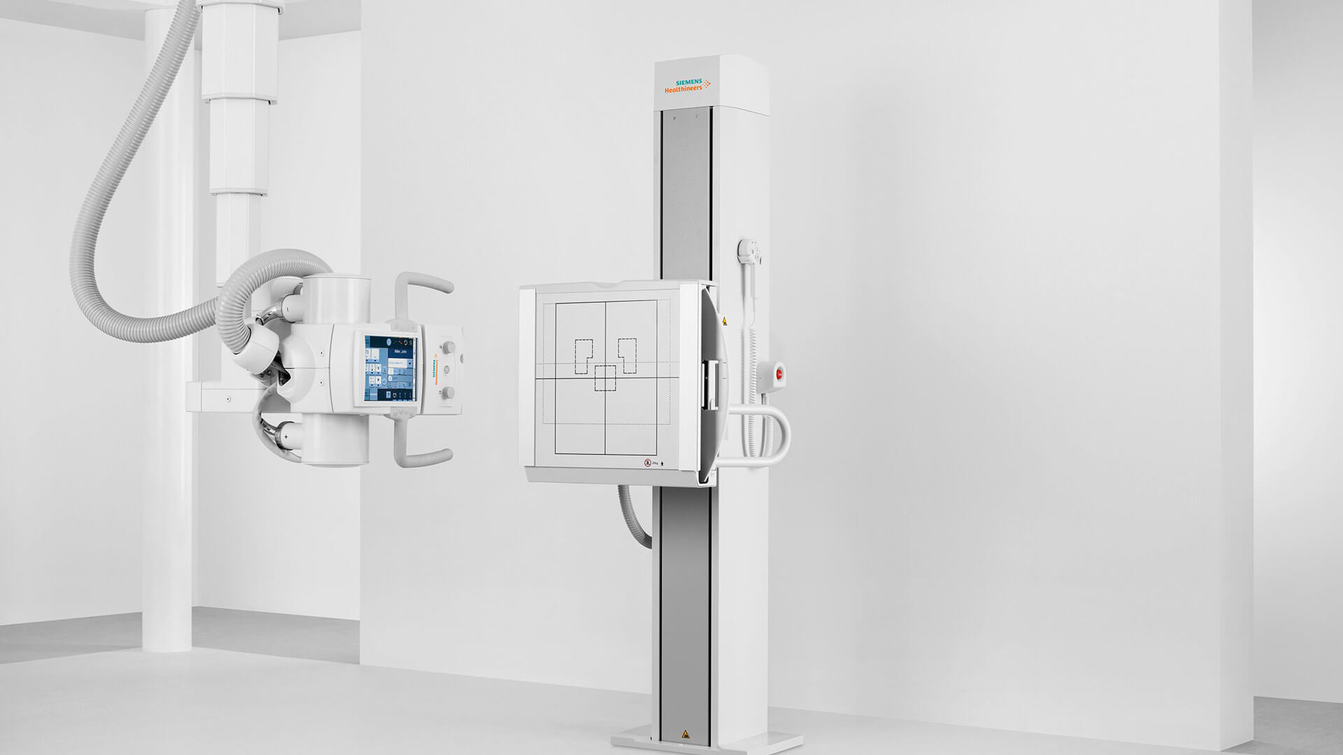
How is an X-ray performed?
The procedures associated with an X-ray depend on the anatomical area being evaluated, but in general, the patient is positioned on a table or against a flat surface, depending on the body part being examined. Usually, chest X-rays, cervical spine X-rays, and shoulder X-rays are performed while the patient is standing, while pelvic and lower limb X-rays are performed with the patient lying down. X-rays of the elbow, forearm, or hand are taken with the patient seated.
Next, the X-ray equipment is directed toward the area to be evaluated and operated by a technician. The technician may produce images at different angles, and it is essential for the patient to remain still to obtain the clearest image possible. Sometimes, the patient may be asked to hold their breath at certain moments to avoid compromising the image. The exam is painless and takes only a few minutes.
Precautions for X-rays
Generally, no special preparation is required before having an X-ray. Patients can eat, drink, and continue taking their regular medications. However, in the case of contrast X-rays, it may be necessary to suspend the use of certain medications or fast for a few hours. The doctor will provide appropriate instructions based on the type of X-ray being performed.
On the day of the exam, it is advised that the patient wear loose and comfortable clothing. Patients will be asked to remove clothing and accessories containing metal, such as garments with zippers, earrings, or watches, as they interfere with the passage of radiation.
As a precaution, patients may need to wear specific protection to reduce their exposure to radiation during the exam. However, X-rays are generally safe since they emit minimal radiation. They would only be harmful if they occurred at high and repeated radiation doses.
Nevertheless, X-rays are not recommended for pregnant women or women suspected of being pregnant unless it is an emergency. It is up to the doctor to carefully assess the risks and benefits to decide if the exam is viable.
X-rays at Joaquim Chaves Saúde
The Imaging units of Joaquim Chaves Saúde medical clinics are a national reference for all types of imaging exams. We have the most advanced technological means for this type of diagnosis, operated by specialized and experienced teams. Take advantage of the most advanced protocols in X-ray exams and make your appointment now.