Mammography is one of the main tools for preventing breast cancer. Discover everything about this exam that can save lives.
In Portugal, 7,000 new cases of breast cancer arise every year. Mammography allows for early diagnosis and saving lives. Find out how it's done, when to have it, and what precautions to take.
What is mammography?
Mammography is an imaging exam that uses low-dose X-ray radiation to analyze breast tissue. The radiation used is low and has no known side effects.
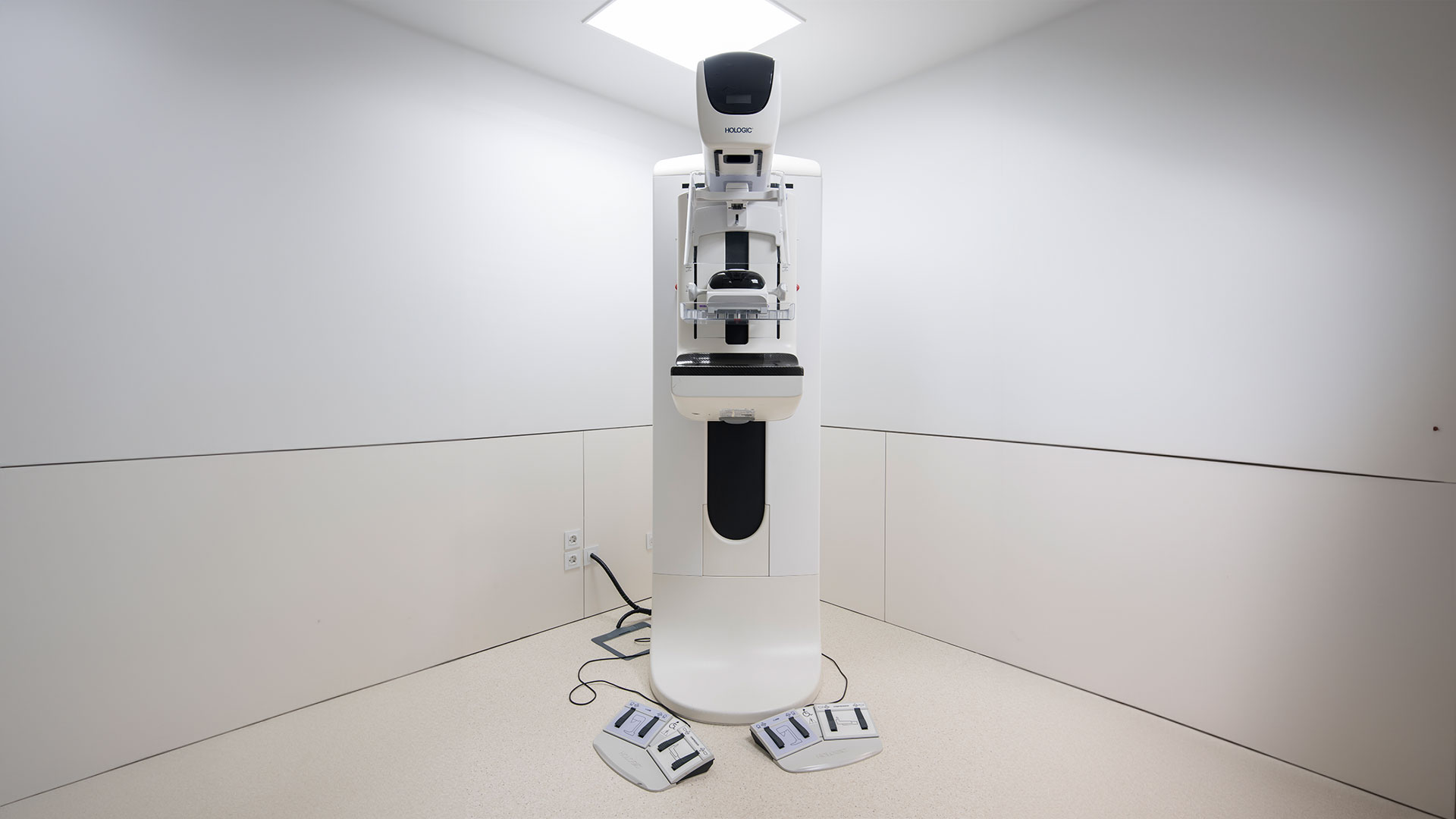
This exam is performed using a device called a mammography machine, which detects changes in the breast, such as nodules or clusters of microcalcifications. Mammography enables the identification and distinction of various conditions, including benign and malignant tumors, with great early detection and accuracy.
What is mammography used for?
Mammography can be used as a breast cancer screening tool, always in conjunction with breast ultrasound. This means that it allows for the detection of breast cancer even before the woman or the doctor identifies any changes through breast palpation. When performed regularly as recommended, it significantly reduces mortality and enables curative treatment of breast cancer.
Therefore, mammography is an essential tool in the fight against breast cancer, crucial for early diagnosis and the cure of a high percentage of cases. Thus, together with their doctor, women should take a proactive role in prevention, integrating mammography into their surveillance routine.
It is also possible to undergo mammography outside of screening if there is suspicion during breast palpation or other performed exams.
When should you have a mammography?
Since the goal of mammography is to screen for breast cancer, this exam should be performed even in the absence of symptoms. The recommendations from the General Directorate of Health for breast cancer surveillance are as follows:
- From the age of 20: monthly breast self-examination, after the menstrual period;
- From the age of 35: mammography every 18 months;
- After menopause: mammography every 24 months.
The doctor may adjust these intervals based on personal or family risk factors. Mammography is not recommended for men. However, since men can also develop breast cancer, they should undergo this exam in the presence of symptoms such as the appearance of a lump.
How is mammography performed?
The performance of a mammography is preceded by an interview where the technician explains the procedure and answers any questions. Then you will be asked to lean against the equipment so that the technician can properly position the breast. The equipment compresses each breast horizontally and vertically, which may cause some discomfort. However, compression is absolutely necessary to obtain a high-quality and conclusive image. Otherwise, breast tissue can overlap and compromise the exam's result.
However, according to international standards, breast cancer screening includes mandatory breast ultrasound, which increases screening accuracy by identifying a higher percentage of tumors.
When the breast is denser, changes may be more difficult to identify. In the presence of suspicious signs, a breast biopsy may be indicated, preferably on the same day. This reduces the waiting time for diagnosis, which can cause anxiety. Early diagnosis also allows for faster treatment initiation, ensuring the best results. The biopsy is performed under local anesthesia, with the aim of collecting samples from the breast lesion.
The samples are analyzed in the laboratory, and a definitive diagnosis is made, without which no treatment should be performed.
What types of mammography are there?
Generally, mammography is performed on each breast (bilateral mammography), but it can also be done on only one breast (unilateral mammography). This is the case, for example, when evaluating mastectomized patients or when an additional view is needed to improve diagnostic sensitivity. In addition, there are two types of mammography, which we will describe below.
Digital mammography
Digital mammography captures images of the breast in a similar way to digital cameras. In this method, the technician can manipulate the digital image to obtain the best possible visualization, zooming in on details or adjusting characteristics such as brightness and contrast. Moreover, the computer itself helps to search for areas of abnormal density, masses, or calcifications that may indicate the presence of cancer.
3D mammography
3D mammography, also known as breast tomosynthesis, represents an important technological advancement in radiology. Through this method, multiple different planes of the breast are captured to form a final three-dimensional image. As a result, the interpretation is more comprehensive, and the diagnostic accuracy is higher. This method of mammography is increasingly used as a screening exam.
What are the risks of mammography?
Mammography does not pose any risks to women. The radiation dose used is minimal and far outweighs the associated risks. However, depending on individual sensitivity, the exam may be
uncomfortable due to breast compression. When performed by competent and experienced technicians, it does not cause pain.
What precautions should be taken before having a mammography?
No prior preparation is required for the exam. However, there are some important factors to consider to ensure that the exam proceeds as smoothly as possible. Firstly, do not undergo mammography while menstruating or if you experience breast tenderness during this period. Ideally, mammography should be scheduled for the week following the menstrual period. The same applies if you are in the premenopausal phase with increased sensitivity to pain.
At the time of the exam, you should have the medical prescription and any previous screening exams (mammography and breast ultrasound), regardless of the date or location of the exams. You should inform the technician of any changes you have noticed in your breast, any previous surgeries, ongoing or past hormonal therapy, and personal or family history of breast cancer.
You may be asked to remove clothing and accessories that interfere with image acquisition, such as earrings, rings, necklaces, or bracelets. You should remain as calm as possible and follow the technician's instructions to ensure a quick and efficient exam. It is important to remember that mammography cannot be performed on pregnant women due to the use of ionizing radiation. If there is a possibility of pregnancy, you should inform the doctor.
Mammography Results: What are the possibilities?
After the mammography is performed, the results are interpreted and classified. The classification used is defined by the American College of Radiology (ACR) and is called BI-RADS (Breast Imaging Reporting and Data System).
- BI-RADS 0: Additional mammography is required because the exam was inconclusive.
- BI-RADS 1: Normal result.
- BI-RADS 2: Detected benign changes, not suspicious.
- BI-RADS 3: Detected benign changes that require further evaluation.
- BI-RADS 4 and 5: Additional evaluation required through biopsy.
- BI-RADS 6: Lesion already diagnosed as breast cancer, biopsy result is always necessary.
Joaquim Chaves Saúde, a leader in diagnostic imaging
Joaquim Chaves Saúde medical clinics are equipped with state-of-the-art diagnostic equipment, such as the 3D mammography machine, which uses the latest image reconstruction algorithms. The results of mammography exams can be obtained quickly and with high precision, eliminating the anxiety of waiting and uncertainty associated with this exam.
Count on Joaquim Chaves Saúde to benefit from the latest means of diagnosis with cutting-edge technology.
